Review Of Roaring And Possible New Surgical Procedure


Roaring in horses is not just a loud, raspy noise made during exercise; it is an issue in the upper airway that restricts airflow and can limit performance in large breed horses such as Standardbreds, Thoroughbreds and warmbloods. Ontario Veterinary College associate professor of Large Animal Surgery, Taralyn McCarrel, is seeking to develop a surgical procedure that could revolutionize the treatment of tie-back surgery, offering new hope for affected horses.
Roaring, or recurrent laryngeal neuropathy, is a condition where one of the nerves controlling the muscles of the throat becomes dysfunctional. This commonly results in paralysis of the left arytenoid cartilage.
McCarrel provided a quick anatomy lesson, comparing the trachea to a castle: “The airway is made-up of two cartilages called the arytenoids. The arytenoids close like doors to cover the trachea, and then the epiglottis is like the drawbridge that comes up and covers them.” With recurrent laryngeal neuropathy, the control to open the doors is abnormal and overwhelmingly, it is most common that the left side becomes affected.
When the nerve fails, the muscle atrophies and the horse struggles to open the left door of the airway because it lacks the stimulus to contract. Due to the inability for the left arytenoid to be held open, the tissue is left flapping and causes the ‘roaring’ noise. The laryngeal neuropathy puts a limitation on the volume of air available for oxygen exchange which can significantly impact the horse's exercise tolerance, causing them to tire quickly.
McCarrel pointed out, “This condition is long recognized, with scientific reports dating back to before the formal establishment of veterinary medicine.” Affected horses often exhibit signs of nerve abnormalities throughout their bodies, though these do not have observable consequences aside from the paralysis of important laryngeal muscles. The most noticeable sign is the loud noise produced during exercise if there is a limitation in airflow.
Prevalence and affected breeds
Roaring is most common in large breed horses, with draught horses estimated around 33 per cent prevalence. Thoroughbreds have a lower prevalence, ranging from two per cent to eight per cent, while warmbloods are also affected. The condition is often detected early in racehorses due to routine scoping, but in warmbloods, it may go unnoticed until they are older and working harder. Interestingly, some high-level dressage horses can have a completely paralyzed arytenoid cartilage yet make minimal noise, possibly due to head position, speculated McCarrel.
Tie back surgery
Prosthetic laryngoplasty, commonly known as tie back surgery, is the current gold standard for treating roaring. This involves making an incision under the horse's neck and suturing the paralyzed cartilage to keep the airway open. Incisional healing typically takes 10-14 days. McCarrel recommends at 30 days post-surgery the horse is scoped to ensure proper healing.
Some surgeons recommend waiting six weeks for the position of the arytenoid cartilage to stabilise following tie back surgery. McCarrel explained, “We try and put the cartilage in a certain open position but there's a strong tendency for it to collapse back to some degree and there's some reset. Most horses will settle at their final position around six weeks.”
Success rates vary widely in literature with non-racehorses generally having a better prognosis due to their lower dependence on maximum sprint-level athletic effort.
Limitations of the current tie-back procedure and new ideas
The tie-back procedure, performed to improve airflow in horses with one-sided laryngeal paralysis, has shown varying outcomes that may be influenced by the surgeon's skill. While the complication rate is not extremely high, searching for new ways to minimize complications and optimize results increases the likelihood of horses returning to optimal performance.
One of the most common complications is the arytenoid cartilage not staying as open as intended. McCarrel said, “If we model airflow, then in theory we need to get 88 per cent of maximum to be able to return airflow to be similar to normal.”
Many horses will fall below this threshold, raising questions if they can reach their full competition or racing potential.
The current tie-back procedure involves sutures that can stretch, slip or cut into the cartilage (causing a loose loop), leading to inconsistent results. “The back of the cartilage we tie the arytenoid cartilage flap to is very thin,” said McCarrel citing another challenge of the current surgical approach.
It was while looking at a CT (computed tomographic image) of a skull, that went as far back as the larynx, that inspiration hit McCarrel. Eyeing up the cricoid, which has thicker cartilage where it contacts the arytenoid cartilage, McCarrel conceived a surgical method that could do away with the knot and sutures and instead use a screw which could result in better stability.
McCarrel also saw the potential for this new surgery to become minimally invasive, only requiring a tiny incision, like what is used in arthroscopy. This would minimize soft tissue trauma that can interfere with the lining of the esophagus, which most often occurs when placing sutures. “If we can place this screw through a little stab incision, we can potentially avoid impinging on and potentially damaging the muscles and nerves in that area,” hypothesized McCarrel. This could reduce some of the less common complications of tie back surgery like chronic cough or dysphasia (feed entering airways), abnormal swallowing leading to lower airway contamination and reflux of saliva from the esophagus.
Step one: The initial step, already published, demonstrated the ability to CT the equine throat and produce models for measuring the desired parameters. The dimensions of pins and screws were also modeled to ensure they could fit across the targeted area.
Step two: Phase two was performed by McCarrel’s former resident creating models with a screw rather than suture holding arytenoid cartilage open to the thicker cricoid cartilage. It was then exposed to negative pressures in an airflow chamber for basic proof of concept.
Next steps and use of CT
The next step, funded by Equine Guelph, aims to develop a minimally invasive surgery for positioning cartilage through a tiny incision. The current project involves modeling the shape and size of horse airways to create inserts that fit over standard intubation tubes. Via the mouth, these inserts will push the arytenoid cartilage into the desired position, as confirmed on CT prior to surgery.
This requires CT scans of many specimens to create models and determine the number of different inserts needed. The goal is to select the appropriate size insert for each horse during surgery, ensuring precise abduction and opening.
Future steps will include developing an approach and guide to pre-plan screw size and exact placement to ensure accuracy and avoid going into the airway.
Introducing CT (computed tomographic) 3-D guidance will be a key component to the development of a less invasive procedure. “We can do all the planning before we make the incision,” said McCarrel, “so when it comes time to do the actual surgery, the surgical approach will be very small, and the surgical time will be short.”
“For years now we've had this move in human surgery to increase the amount of imaging guidance in order to have smaller incisions, more precise and minimally invasive surgery,” said McCarrel.
This new approach aims to provide more accurate implant placement with less disruption of muscles and nerves in the larynx, minimize complications and improve outcomes for horses with laryngeal paralysis.
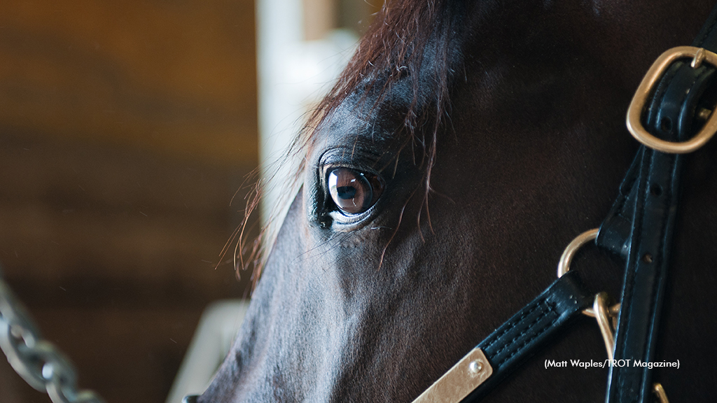
(Equine Guelph; Racehorse pictured does not have laryngeal neuropathy)